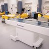
When John Carroll University students begin their 2026-27 academic year in earnest this Fall, they’ll have the opportunity to train and learn on a piece of equipment used by many of the leading health science schools across the country: the Anatomage Table, a state-of-the-art 3D digital life-size virtual dissection table.
The arrival of the table underscores JCU’s growing academic distinction in pre-medical, nursing, and health science education, and was made possible through extraordinary leadership support from the brothers of the Iota Chi Upsilon fraternity, and generous contributions from across the JCU community.
The cadaver CT scan images provide fully interactive renderings, allowing students in human anatomy and physiology labs and nursing courses to trace intricate vascular networks, observe cardiac motion in real time, inspect more than 1,100 digital histology scans, and practice specialized procedures such as ultrasound and catheterization with high-precision visualization.
"The Anatomage Table delivers ultra-high-quality, life-size 3D renderings of real human anatomy right to our fingertips," said Melissa Cole, Dean of the College of Health. "Our students can look forward to slicing, rotating, and examining complex structures with a level of detail and flexibility that traditional labs simply can't match. This table will greatly expand opportunities for hands-on learning, and elevate learning outcomes for our students.”
To that end, studies have shown that by incorporating the Anatomage table into anatomy & physiology curricula can improve test scores by up to 27%.This ability to interact with real human structures and live physiological systems allows students to build deep conceptual mastery before they step into clinical practice ensuring JCU graduates enter the healthcare workforce with a distinct competitive advantage.
For faculty, the table deepens the academic rigor of the health sciences curriculum.
"This is a really great opportunity for our students," said Carol Mack, Exercise Physiology and Biomechanics Lab Coordinator at JCU. "They can learn actual human structures in 3D through this table, and really for anyone who's going into healthcare or exercise professions, they need this knowledge."
Beyond foundational anatomy, the table allows instructors to bridge the gap between structure and human movement. By linking muscle function, vascular pathways, and live ECG readings, faculty can walk students through complex physiological responses as they happen.
"When we need to understand what we're testing with certain muscles, now they're actually going to have an understanding of where that muscle is, how it functions, and how the blood supply goes to it," Mack added. "Seeing how the heart is beating through the ECG simulations—it's a game changer for us."